Monday 31 August 2015

Mitochondria


Mitochondria are commonly known as power house of the cell ( because it take part in release of energy in the form of ATP by oxidative phosphorylation ). They were first observed by Kolliker in 1850. Benda (1897) gave the present name of mitochondria.
                                                                            Mitochondria are absent in prokaryotes or anaerobic eukaryotes. Their number varies from one in some algae ( Chlorela ), 25 in sperm cell, 300-400 in kidney cell, 1000 in liver cell, 50,000 in giant amoeba and 500,000 in flight muscle cell. The number depends upon cellular activity. They are different in shape - sherical, cylindrical, tubular or filamentous.

Ultrastructure:- The mitochondria contains two membranes and two chambers, outer and in inner. The two membranes form the envelop of the mitochondria.

Outer Membrane:- The membrane is smooth. It is permeable to number of metabolites. It is due to presence of protein channels called porins. A few enzymes connected with lipid synthesis are located in the membrane.
Inner Membrane:- It is permeable to some of metabolites. It is rich in double phospholipids. Protein content is also high. The inner membrane is infolded variously to form involutions called cristae. They are meant for increasing the physiological active area of inner membrane. A cristae encloses a space that is continuation of the outer chamber. The inner membrane as well as cristae posses small tennis-racket like particles called elementary particles, F- Fparticles or oxysomes. Each elementary particle or oxysome has a head, a stalk and a base. Elemetary particles or oxysomes contain ATPase enzyme and are connected with ATP formation. They are, therfore, the centres of ATP synthesis.


Outer Chamber:- The chamber is the space that lies between the outer and inner membrane of the mitochondrial envelop. It extends to into the spaces of cristae. The chamber contain a fluid havin a few enzymes.
Inner Chamber:- It forms the core of the mitochondria. The inner membrane contains a semi-fluid matrix. The matrix has proteins, ribosomes, RNA, DNA, enzymes of Kreb's cycle, amino acid synthesis and fatty acid metabolism. Mitochondrial ribosomes are 55s to 70s in nature. They thus resemble to the ribosomes of prokaryotes. Because of presence of its own DNA this organelle is semi-autonomous.


Active and inactive state:- Mitochondria occur in two states, active and inactive. In active state, mitochondria are actively engaged in performing Kreb's cycle, electron transport chain and oxidative phosphorylation. In this state core is reduced, cristae randomly distributed and outer chamber space quite large. Active state is also called condensed state. In inactive state, Atp synthesis is reduced. Matrix or core is enlarged while outer chamber is narrow. It is also called orthodox state.

Functions of Mitochondria:-
i) Mitochondria are miniature factories where respiratory substrate is completely oxidised to carbon dioxide and water.The energy is liberated in the form of ATP. ATP comes put of mitochondria and helps perform verious energy requiring processes of the cell.Because of formation of ATP the mitochondria is called power houses of the cell. 
ii) Mitochondria provide important intermediates for the synthesis of biochemicals like chlorophyll, cytochromes, steroids etc.
iii) The inner chamber of the mitochondria has enzymes for the synthesis of fatty acids.
iv) Synthesis of many amino acids occur in the mitochondria.
v) Mitochondria may store and releases calcium when required.




Thursday 27 August 2015

Cell Wall

The cell wall is secreted by the protoplasm. It was first observed by Robert Hooke in 1665 in a thin slice of cork.
              The cell wall is chiefly composed of insoluble polysaccaride, cellulose, certain other compound such as hemicellulose, pectin, lignin, suberin, xylan and silicates of calcium and magnesium also occur in the cell wall.

Structure:- the cell wall is composed of four layers. a) middle lamella b) primary wall c) secondary wall and d) tertiary wall
a) Middle lamella:- It is the first formed wall layer, formed at the time of cytokinesis ( division of cytoplasm is called cytokinesis ). It is made up of Ca and Mg pectate. It is common to all the cells, popularly known as the cementing layer.The softening of ripe fruits is caused by partial solubilisation of pectic compounds to produce jelly like consistency.
b) Primary wall:- It is laid down along the middle lamella. It is made up of cellulose and pectin and is about 1-3 micro meter in thickness. Meristematic ( those cells who can divide are called meristematic cells in plants ) and parenchyma cells have primary cell wall only.
c) Secondary wall:- It is thick and rigid and laid down next to the primary wall. It again consists of three sub-layers called S1, S2 and S3. It is made up of cellulose, lignin or suberin. It develops in those cells which cease to grow e.g. in tracheary elements and sclerenchyma.
d) Tertiary wall:- internal to secondary wall is another wall material. It is chiefly composed of xylans. this is found in the tracheids of gymnosperms.

Functions of cell wall:- i) It maintains the shape of the cell.
ii) It protects the cell from mechanical injury.
iii) It allows the material to pass in and out of the cell.
iv) Many enzymatic activities are known to occur in the cell wall.
v) Cutin and suberin check loss of water by transpiration.
vi) It provides mechanical support aganist gravity. It is due to the rigid cell wall that the aerial parts of the plants are able to keep erect and expose their leaves to sunlight.
vii) It counteracts osmotic pressure.


Diffusion

The movement of molecules, atoms, ions of gases, liquid or solids from region of higher concentration to region of lower concentration until the molecules get uniformly distributed irrespective of the law of gravitation.

Diffusion Pressure:- It can be defined as the potential ability of a molecule or ions ( solid, liquid or gas ) to diffuse from the area of greater concentration to an area of lesser concentration. The diffusion pressure is directly proportional to the concentration or number of diffusing particles.

Factors affecting diffusion:- i) Rate of diffusion is directly proportional to the temperature.
ii) The rate of diffusion is inversely proportional to sequare root of the density of the diffusing substance.  
iii) The rate of diffusion is directly proportional to the surface area from which diffusion occurs.
iv) Rate of diffusion is directly proportional to the difference of diffusion pressure at the two ends.

Importance:- i) The exchange of gases e.g CO2 intake and O2 output in photosynthesis and CO2 output or O2 intake in respiration takes place by the principle of independent diffusion.
ii) The process of diffusion involved in the transpiration of water vapours.
iii) The ions are absorbed by the simple diffusion during passive salt intake.
iv) Diffusion is an effective means of transport of substances over a very short distances.

Imbibition

Imbibition is the phenomena of adsorption of water or any other liquid by the solid particles of a substance without forming a solution. The solid particles which adsorb water are called imbibants. The imbibants are rich in hydrophilic colloids and have strong affinity with water.
                                                                                     Imbibition results in as increase in volume, liberation of heat and development of pressure called imbibition pressure.

Importance:- i) Absorption of water by young cells is mostly through imbibition.
ii) Imbibition plays important role in germination of seeds.
iii) Imbibition causes the swelling of seeds results in the breaking of seed coat.
iv) Seedling is able to come out of soil due to development of imbibition pressure.
v) Jamming of wooden frames during rains is caused by swelling of wood due to imbibition.
vi) In older times, it was used in breaking the rocks.

Factors affecting imbibition:-
i)  The rate of imbibition increases with rise in temperature.
ii) The rate of imbibition decreases with increasing the concentration of solutes in medium.
iii) Imbibition also influenced by texture like more looseness more imbibition and compactness less imbibition.

Imbibition pressure:- The imbibant, after imbibition of water or liquid, exerts the pressure which is called imbibition pressure. The germinating seeds have imbibition pressure of about 1000 aiomspheres.

Plant-Water Relations

Water is one of the most important constituent of the protoplasm and body fluids. It forms 50 - 90% of the total body weight of living beings and is the universal solvent.
                                                                            Gases, minerals and other solutes enter the plants and move from cell to cell dissolved in water. It provides turgidity to growing cells which is essential for plants to maintain their form and structure. It helps in regulating the body temperature.

Osmoregulation in plants:- Osmoregulation is the homeostatic process in which organisms maintain their internal conditions constant or in which organisms maintain the concentration of their body fluids and temperature at a steady state. In plants this is maintained by absorption, transport and retention of water in their bodies. Transpiration ( loss of water in the form of water vapour from the exposed surface of the plant ) results in the loss of water by plants but this loss is made up by absorption of water.
                The forces like imbibition, diffusion and osmosis are responsible for exchange of material between cells and environment and between cells.

Tuesday 25 August 2015

Membrane Transport

Passage of substances across biomembranes occur by three methods -
1) Passive transport
2) Active transport
3) Bulk transport

1) Passive transport:- It is the mode of membrane transport in which cell does not spend any energy. it is of two types:-
a) Passive diffusion by tunnel protein molecules:- The tunnel protein molecules have the channels. these channels permit water and water soluble substances to pass through them. The channels only permit the molecules to pas from higher concentration to lower concentration gradient.
b) passive transport by carrier proteins:- The proteins are known as permeases. They transport the material down the concentration gradient. A carrier protein combines with a specific substances to be transported and moves it from one side of the membrane to another through a channel in it.
                                                             Facilitated diffusion moves glucose molecules in liver and it occurs without expenditure of energy.

2) Active transport:- Active transport moves substances against the concentration by utilizing energy which is privided by ATP ( Adenosine triphosphate ). The carries protein has a binding site for ATP in addition to the binding site for the substrate. ATP molecules binds to the carrier protein. The energy set free brings the substrate binding site of the protein to the surface of the membrane. The substrate present in the medium joins the binding site of protein, forming carrier-substrate complex. It undergoes conformational changes and carries the substrate through a channel in it to the cytoplasmic site of the membrane. Here the substrate is released. This type of transport by using energy is supported by various evidences:- i) Absorption is reduced with the decrease in oxygen content of the surrounding environment ( cells are using oxygen in the respiration involved in the liberation of energy-ATP).
ii) Metabolic inhibitors like cyanides inhibit absorption.
iii) Absorption of different substances is selective.
iv) Decrease in temperature decreases absorption. ( again respiration process is regulated by temperature)
v) Active transport is more rapid then diffusion.

3) Bulk transport:- It occurs by two methods:- pinocytosis and phagocytosis. They involve the enclosure of the material under transport in the vesicles of the membrane. The inward transport by means of carrier vesicles is called endocytosis. The outward transport of substance by means of carrier vesicles is known as exocytosis.
Pinocytosis:- It is the bulk trasport of fluid matter and substances dissolved in it. pinocytosis is also called cell drinking.
Phagocytosis:- It is also called cell eating. Phagocytosis is the transport of solid matter like food, foreign particles, pathogens etc. across the membrane by forming vesicles. These vesicles are called phagosomes. phagosomes fuses with lysosomes to produce a digestive vacuole. The solid food is digested. The digested food diffuses into the cytoplasm. The vacuole with indigestible substance is called residual vacuole. The undigested part are usually thrown out of the cell in the process of exocytosis called ephagy or cell vomiting. Phagocytosis by some white blood cells is an important defence mechanism of the animal body.


Function of Cell Membrane

These are the important functions of cell membrane.
1) The cell membrane cause compartmentalisation. As plasma membranes they separates the cell from external environment.
2) Plasma membrane protect the cell from injury.
3) The plasma membrane allow the flow of materials and information between one cell and another, the cell membrane makes metabolism possible.
4) Plasma membranes of the adjacent cells form various types of junctions for keeping the cells together.
5) plasma membranes as well as other membranes of the organelles have selective permeability, they allow only selected substances to pass inwardly. The membranes are impermeable to others.
6) Membranes have carrier proteins for active transport.
It helps certain cells in movement by forming psudopodia as in Amoeba and leucocytes.
7) Substances attached to cell membrane determine antigen specificity.
8) In nerve cells the cell membrane takes part in transmission of impulses.
9) Membrane infolds are used for bulk intake of materials by endocytosis.
10) Cell membranes contain enzymes for performing certain reactions on their surfaces. e.g. ATPase
11) As microvilli the membrane becomes specialized for the absorption of substances.

Different types of membranes:-
a) Permeable membrane :- These membranes allow all types of solute, solvent and ions through them e.g. cellulose wall of cells.
b) Impermeable membrane :- These types of membranes does not allow the diffusion of both solute and solvent molecules. e.g. heavily cutinised cell walls in plants.
c) Semipermeable membranes :- These types of membranes allow diffusion of solvent molecules but do not allow the passage of solute particles. e.g. parchment paper.
d) Differentially permeable membranes :- These are also called selectively permeable membranes. These type of mmbranes allow only selected solute molecules and slovent molecules to pass through them. e.g. plasma membrane.


Sunday 23 August 2015

Cell Membrane

The term was originally used by Nageli and Cramer (1855) for the membranous covering of the protoplast. Plasmalemma or plasma membrane was discovered by Schwann (1838). Membranes also occur inside the cytoplasm of eucaryotic cells as covering of several organelles like nucleus, mitochondria, plastids, lysosomes, Golgi bodies etc. Vacuoles are separated from cytoplasm by a membrane called tonoplast.
All membranes whether external or internal are now called biomembranes. Average thickness is 75 Angstrom. Biomembranes are selectively permeable membranes.

Composition:- Chemically biomembrane consists of lipids (20-79%), proteins (20-70%), carbohydrates (1-5%) and water.The important lipids of the membrane are phospholipids, sterols, glycolipids, sphingolipids.
                                     The lipid molecules are amphiatic or amphipathic, they posses both polar hydroplilic (water loving) and nonpolar hydrophobic (water repelling) ends. The hydrophilic region is in the form of head and hydrophobic tails usually occurs towards the center of the membrane. It results in the formation of lipid bilayer.
                                                   Sveral types of models have been put forward to explain the structure of biomembrane.
Lamellar Models :- They are the early molecular models of biomembrane. according to these models biomembranes are stable layered structures.
Danielli and Davson Model :- This model can be described in the following points:-

1) According to this model the plasma membrane consists of a biomolecular layer of phospholipids which is sandwiched between the two layers of proteins.
2) The molecules of phospholipids are arranged in the two parallel chains.
3) The polar hydrophilic heads of the phospholipid molecules are facing towards outer surface while hydrophilic tails lie toward the centre.
4) The protein layers are asymmetrical. On one side folded beta chain proteins are present while on the other side globular proteins are present.
Drawbacks :- a) this model fail to explain how the water and water soluble materials pass through cell membrane.
b) How the biomembranes are flexible.
c) How active transportation occurs through biomembranes.


Robertson Model :- J. David Robertson (1959) modified the model of Danielli and Davson. This model can be described in the following points:- 
a) The cell membrane consists of three layers - outer dark, middle light and inner dark. The outer and inner proteins layers and middle one is layer of phospholipids. 
b) According to this theory the cell membrane is a unit membrane, consisting of a biomolecular lipid layer between the two protein layers.


                                                    Robertson Model of cell membrane


c) Each protin layer is 20 angtsrom in thickness and the bimolecular lipid layer is 35 angstrom. 
                      It is simply the modification of sandwich model therefor it faces the same drawbacks as of sandwich hypothesis. 

Mosaic Model :- It is most recent model of biomembrane proposed by Singer and Nicolson in 1972.
According to this model the membrane dors not have a uniform disposition of lipids and proteins. Further the membrane is not solid but is quasifluid. 
                                      Fluid-mosaic model postulates that the lipid molecules are present in a viscous bilayer as in lipid layer. Protein molecules occur at place both inside and on the outer side of lipid bilayer --  Proteins icebergs in a sea of lipids. The internal proteins are called intrinsic or integral proteins while the external ones are known as extrinsic or peripheral proteins.The intrinsic proteins account for 70% of the total membrane proteins. Some of the intrinsic proteins run throughout the lipid bilayer. They are called tunnel proteins. The extrinsic proteins are present superficially on the two surfaces of the membrane. Many membranous proteins function as enzymes. Some of them behaves as permeases. Some lipids on the outer side possess small carbohydrate molecules to form glycolipids. 

Evidences in support of Fluid Mosaic Model:- 
1) The model provides the occurrence of protein particles both on the surface and interior of cell membranes. 
2) Fluid mosaic model can explain the presence of different types of permeability of cell membrane.
3) It accounts for dynamic nature of biomembranes.
4) The model explain the passage of both electrolytes and non-electrolytes.


                                       Fluid-mosaic model of a membrane


Friday 21 August 2015

Eucaryotic cell

A cell is an organised mass of protoplasm surrounded by a protective and selectively permeable membrane. protoplasm of cell is called protoplast. It is made up of cytoplasm, nucleus and vacuoles. Electron microscope has shown that cytoplasm has a complex organisation formed of cytoplasmic matrix and cell organelles. Genetic material or DNA is organised into chromosomes and chromotin.
                                      In the cytoplasm matrix large number of cell organelles like endoplasmic reticulum, plastids, mitochondria, Golgi bodies, centrioles, lysosomes, sphaerosomes, peroxisomes, vacuoles etc. are present. doubling membrane coverings occurs around plastids and mitochondria. Single membrane covering is found over endoplasmoc reticulum, Golgi bodies, lysosomes, sphaerosomes, peroxisomes and vacuoles. Organelles without a membrane covering are ribosomes.

Different types of cells in multicelluar organisms

A multicellular organism is composed of numerous cells. The cells are of three main types:-

1) Undifferentiated cells:- Undifferentiated cells and stem cells have power of division. they are unspecialised cells and they divide to form new cells e.g. stem apical meristem , root apical meristem, vascular cambium, cork cambium, germinal epithelium, bone marrow etc. Zygote is an also undifferentiated cell. Basically these cells helps in growth of an organism.

2) Differentiated cells:- These cells are specialized to perform particular function. These cells have lost the power of division. They are derived from the undifferentiated cells. Differentiation leads to better organisation, division of labour ( it is the typical character of multicellular organisms in which group of cells perform one particular function this group is called tissue ) and higher efficiency.

3) Dedifferentiated cells:- These cells are derived from differentiated cells. Under normal condition differentiated cells fail to divide, but under exceptional circumstances they start dividing. the process by which they lose their specialization is called dedifferentiation. Cork cambium of plants is always produced through dedifferentiation.  

Tuesday 18 August 2015

Components of Bacterial Cell

A bacterial cell consists of a cell envelop, cytoplasm, nucleoid, plasmids, inclusion bodies, flagella, pili and fimbriae.

Cell envelop :- It is the outer covering of protoplasm of bacterial cell. Cell envelop consists of three components- glycocalyx, cell wall, cell membrane.
Glycocalyx :- ( Mucilage sheath) It is the outermost mucilage layer of the cell envelop occurs in the form of loose sheath also called slime layer. It has several secondary functions:- a) Prevention of desiccation.   b) Protection from phagocytes.  c) Protection from toxic chemicals and drugs.   e) Attachment.   f) Immunogenicity.
Cell Wall :- The cell wall is a rigid and made up of heteropolymer mucopeptide complex containing N-acetyl glucose amine and N-acetyl muramic acid and peptidoglycans. They are cross linked by small peptides chains. Lysozyme the antibacterial enzyme of tears, saliva, gastric juices etc. has the ability to hydrolyse the peptidoglycan. 
Plasma Membrane:- It is selectively permeable membrane that forms the inner most layer of the cell envelop. Bacterial plasma membrane has a structure similar to typical membrane. It is made up of phospholipids  and proteins and some polysaccharides . 
Cytoplasm:- It is a colourless, semi-fluid material which is composed of proteins, lipids, carbohydrates, water and minerals salts. Membrane bound cell organelles as found in eukaryotes are absent. Cytoplasmic streaming is absent. Sap vacuoles are absent. Various structures are found in cytoplasm are as follows;-
i) Mesosome:- The membrane is inverted at certain places in the cytoplasm. These invaginations are known as mesosome. Mesosome are of two types-  septal and lateral. Septal mesosome connects nucleoid with plasma membrane and helps in the cell division. Lateral mesosome are not connected with nucleoid. It contains respiratory enzymes therefor , also called chondrioid.
ii) Ribosomes:- They are small membraneless, submicroscopic ribonucleoprotein structures. The ribosomes are 70S in nature ( S denotes sedimentation coefficient or Svedberg number). Each ribosomes has two subunits, larger 50S and smaller 30S.  Ribosomes are of two types. Fixed and free. Ribosomes generally occur in helical groups called polysomes and polyribosomes.
iii) Chromatophores:- They are internal membrane systems present in photosynthetic prokaryotes. Chromatophores develop as membrane lined sacs or thylakoids.
iv) Nucleoid;- It represents the genetic material of prokaryotes. It is also known as the prochromosome, genophore and chromoneme. Nucleoid consists of a single circular strand of DNA duplex. DNA is naked because of its non-association with histone proteins. Nuclear membrane is absent and nucleoid is embedded freely in the cytoplasm. It is equivalent to the single chromosome of eukaryotes.
v) Plasmids:- They are self replicating, extra chromosomal segments of double stranded, circular, naked DNA. They are independent of main nucleoid. Some of them contains important genes like fertility factor, nif genes and resistant factor.  Plasmids which can get associated temporarily with nucleoid are known as episomes.
vi) Inclusion bodies:- They are non-living structures present in the cytoplasm. These are gas vacuoles, inorganic inclusions and food reserve. Gas vacuoles are gas storing vacuoles found in cyanobacteria, purple and green bacteria. Inorganic inclusions are sulphur granules, iron granules and magnetite granules. Food reserv are cyanophycean starch, lipid globules and protein granules.
vii) Flagella:- A bacterium flagellum is a long whip like structure projecting on the surface. It is formed of single fibril. It arises from a basal plate situated just inside the plasma membrane. The flagella helps in locomotion. The flagella is made up of flagellin protein.
viii) Pili:- These are small, non-motile hair like structures present on the surface of some forms of bacreia. They help in attachment of bacterial cell, and play important role in bacterial conjugation.


                            Bacteria cell under electron microscope


Structure of a Bacterial Cell

The bacteria are unicellular organisms devoid of chlorophyll which reproduce by fission. The name "bacteria" was first used by Ehrenberg (1838) but they were first observed by A.Von. Leeuwenhock (1938) who named them as tiny animalcules.

Occurrence:- Bacteria can grow in all possible environment i.e. in soil, water, in animals and plant bodies. Thermophilic bacteria can live in hot springs.

Shape and form of bacteria:- In shape bacteria cells are of many types. These are
Coccus:- Coccus bacteria are spherical in outline. Depending on their grouping they are (i) Monococcus (occurring singly), (ii) Diplococcus ( in twos), Streptococcus (in chains).
Bacillus:- The bacterium is straight and cylindrical like a rod.
Spirillum:- The bacterium is coiled like a cork-screw.


Vibrio:- The body of bacterium is like a comma, e.g. Vibrio cholerae
Stalked:- The bacterium possesses a stalk,  e.g. Caulobacter 
Budding:- THe bacterium is swollen at places e,g. Rhodomicrobium.

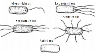
Flagellation:- On the basis of number and forms of flagella there are following types of bacterial cells.
a) Monotrichous:- Single flagellum at one pole e.g. Vibrio.
b) Amphitrichous:- One flagellum at both the ends. e.g. Pseudomonas.
c) Cephalotrichous:-  Two or more flagella at one end only e.g. spirullin midulium.
d) Lophotrichous:- Group of flagella occurs at each of the two ends.e.g. spirilla
e) Peritrichous:- A number of flagella are distributed all over the surface. Bacillus.                                 
f) Atrichous:- Flagella absent.
Gram Positive and Gram Negative Bacteria:- A Danish scholar Christian Gram (1884) on the basis of staining properties classify bacteria into two groups.
                                                               All bacteria turn blue when stained with crystal violet. The bacteria which retain this blue stain after treating with iodine and then with alcohol are called Gram +ve  and those which do not retain the blue stain are called Gram -ve.
Gram -ve bacteria are usually pathogenic and more resistant to antibiotics. e.g. E.Coli, Rhizobium Acetobacter, Azotobacter etc.
Gram +ve bacteria are more susceptible to antibiotics. e.g. Lactobacillus, Clostridium, Bacilli etc.


Thursday 13 August 2015

Prokaryotic cell

The prokaryotic cells are morphologicallly the most primitive cells. A prokaryotic cell is essentially a single envelope system. It is a simple type of cell in which genetic material is not organised in the form of nucleus, it lies freely in the cytoplasm whence it is known as prochromosome. Prokaryotic cells are known for their rapid multiplication. The prokaryotic cells have the following important features:-
1) Nuclear Material:- DNA is naked and lies variously coiled in the cytoplasm. It is often called genophore, nuclear body or nucleoid. Many prokaryotic also have additional small circular DNA called Plasmid.
2) Nuclear component:- Nuclear envelope, nucleoplasm, nucleolus and histone proteins of chromatin are absent.
3) Types:- Prokaryota contains organisms like blue-green algae ( Cyanobacteria, Nostoc ), bacteria, PPLO ( pleuropneumonia  like organism ), archaebacteria, spirochaetes, chlamydiae.
4) Cell wall:- It is present in bacteria and cyanobacteria. Cell wall is absent in mycoplasma or PPLO.
5) Flagella and Fimbriae:- The bacterial flagella are single stranded as compared to 11 stranded in eukaryotes. In some bacteria non-motile appendges called pili or fimbriae also occur.
6) Photosynthetic Thylakoids:- Blue-green algae and some bacteria are photo-autotrophic (who can make their own food with photosynthesis). Their photosynthetic thylakoids lie freely in the cytoplasm.
7) Membrane lined cell organelles:- All double membrane layered organelles are absent like mitochondria. cholroplast, golgi bodies, lysosomes etc.
8) Ribosomes:- Ribosomes are 70s as compared to 80s.
9) One -Envelope System:- There is a single that surrounds the cell. Hence, prokaryotes have a single membrane or one-envelope system.
10) Cyclosis:- Cytoplasm does not show cyclosis or streaming movements ( autonomic vital movements that occurs in cytoplasmic matrix).
11) Spindle:- Mitotic spindle is not formed during cell division.
12) Sexual Reproduction:- It is absent. They multiply very rapidly by asexual means like binary fission, sporulation etc.
12) Transcription and Translation:- Both occurs in cytoplasm ( transcription is a synthesis of RNA over DNA strands and translation is the synthesis of proteins on ribosome)
13) Respiratory Enzymes:- They usually lie in contact with cell membrane.
14) Nitrogen Fixation:- It occurs only in some bacteria and cyanobacteria.


Wednesday 12 August 2015

Cellular Totipotency

Cellular totipotency is the ability of a living somatic cell to form the complete organism. A cell is totipotent because it has a complete genetic information in its nucleus. Somatic cells during their maturation the cells undergo differentiation ( specialized cells for a particular function). Differentiated cells not showing division. Under special circumstances differentiated cells will change into immature cells called dedifferentiation. After dedifferentiation cells will start to multiply.
Totipotency can be easily demonstrated in plant cells.
                                                                                    Cellular totipotency was first proposed by German botanist Haberlandt in 1902. He thought if every somatic cell has the full genetic material then why this cell should not be able to produce whole organism. However he failed to grow new plants from isolated cells of green leaves. He failed due to

1) Lack of aseptic conditions
2) Lack of proper nutrient medium

The two defects removed by White (1932). The scientist successfully grew tomato roots in culture medium. In 1939,  three scientist ( White, Gautheret, Nobecourt ) were independently able to grow callus in tissue culture. Callus is unorganized mass of cells. Skoog and Miller (1957) discovered that callus can be made to develop shoots and roots through changing the ratio of harmones ( IAA and cytokinin). The phenomena is called morphogensis. Cellular totipotency was demonstrate first time by Steward et al ( et al means co-workers) in 1957. they took 2 mg pieces from phloem of carrot roots. These pieces or explants were made up of mature nondividing cells. They placed these explants in liquid culture medium containing coconut milk. There was an active growth and division of cells. After shaking the cells tend to form clusters. Clusters differentiated and formed initials of roots. In a semisolid medium, each developed shoot and gave rise to a plantlets or a small plant.

Advantages:-  1) Quick propagation of useful plants.
                         2) Raising rare plants which are difficult to grow from seeds.
                         3) Multiplying sterile hybrids.
                         4) Rapid production of new verieties.
                         5) Production of virus free plants.
                         6) Production of chemical resistant varieties.
                         7) Reducing the period of formation of new varieties.
                         8) Introducing mutations and selecting mutants.


Cell Theory

   The initial idea of cell theory was given by a French biologist Dutrochet (1824). According to him, the concept that cell is the basic unit of living beings is known as the Cell theory.
    The credit of formulation of cell theory is given to a German botanist M.J. Schleiden and a German zoologist T. Schwann who clearly outline the basic features of cell theory that the cell is the basic unit of life.
Modern Cell Theory ;- In 1858, Rudolph Virchow stated, "Omnis cellula-e-cellula" i.e all living cells arise from pre-existing cells. The old theory was thus revised and the modern cell theory proposed as follows :

1) All living organisms are made up of cells.
2) A cell is a small mass of protoplasm containing a nucleus or nuclear material and some organelles,     bounded by cell membrane.
3) Cells always arise from the pre- existing cells by division.
4) Every organism starts its life from a single cell.
5) The growth of an organism is due to the growth of the cells or due to cell multiplication.
6) All cells are fundamentally similar.
7) All activities of the organism are sum total of activities of different cells.
8) The cells have complete genetic information in genetic material called DNA.

                                              Viruses do not fit in the definition of cell. They don't have their own cellular machinery. they will act like a living cells only by using host cell mechinery or start to behave like a living organism. So outside the living cell Viruses are non-living and inside the body of living organism or cell they are living and starts to multiply.

Tuesday 11 August 2015

Cell - The basic unit of life

Cell is a basic unit of life and in other words it is also a structural and functional unit of life or an organism. It is the smallest unit capable of independent existence and performing the essential functions of life. The organism made up of one cell are called unicellular organisms and the organism consisting of many cells are known as multi-cellular organisms.
Brief History : The study of cell is called as Cytology. The term "cell" was given by Robert Hooke for the first time observed cork cell under the primitive microscope. Anton van Leeuwenhoek observed living cells for the first time. He was the first to discover bacteria, protozoans and other unicellular forms.


                                                           STRUCTURE OF PLANT CELL