Tuesday 29 September 2015

Stomatal Apparatus

Stomata are minute pores present in the epidermis of leaves and other soft parts of the plants which are normally for the gaseous exchange during photosynthesis and evaporation of water during transpiration. In other words stomata are meant for plant to breath. Singular it is stoma is actually the pore surrounded by guard cells ( specialized epidermal cells present with the normal epidermal cells ). They are easily influenced by changing turgor pressure in the guard cells because guard cells are also connected to the adjacent epidermal cells through plasmodesmata. The cells also have chloroplast and vacuole. They can also store starch in few cases.


                                    Gaseous Exchange Through Stomata


In most of the plants the guard cells are kidney shaped and concavo-convex curvature of the guard cells helps in the opening and closing of the pore (stoma) under the influence of changing turgor pressure in guard cells. The walls of these guard cells are thickened on inner side and walls are thinner and more elastic on the outer side. When the guard cell will have full turgor pressure then it swells up on the outer side and inner concave sides also bend out slightly as to create pore. Now the stomata is open. During closing of stomata reverse changes will takes place.


                        .


                                                  Opened and Closed Stomata

In cereals or most of the monocots, the guard cells are dumb-bell shaped in outline.Their extended ends are thin-walled while middle portions are highly thick-walled. In such cases opening and closing of stomatal pore is caused by expansion and contraction of thin-walled ends of the guard cells.


                                                          Dumb-bell Shaped Guard Cells


Thursday 24 September 2015

Factors Affecting Transpiration

External Factors:-
i) Relative Humidity:- Relative humidity is the percentage of water vapours in the air at a given time and temperature. Transpiration is inversely proportional to the relative humidity in the air. If there is more moisture in the air the less amount of water will evaporates from the plant surface.
ii) Temperature:- The rate of transpiration is directly proportional to the atmospheric temperature. If there is increase in the temperature in the atmosphere the humidity will decrease and transpiration will increase. High temperature opens stomata even in darkness hence increases the transpiration.
iii) Light:- Stomata opens in the presence of light and therefore transpiration will takes place. Light increases the rate of transpiration. In other way light also increases the atmospheric temperature and again enhance the rate of transpiration.
iv) Velocity of the Wind:- The rate of transpiration is directly proportional to the velocity of the wind or in other words the movement of air increases the rate of transpiration. A wind velocity of 40-50 km/hr decreases transpiration by closing the stomata due to mechanical effect, drying and cooling of the transpiring organ
v) Atmospheric pressure:- Low atmospheric pressure enhances the rate of transpiration.
vi) Availability of water:- The rate of transpiration is also influenced by the availability of water in the soil. Fully turgid guard cells only responsible for opening of stomata. Therefore, A decrease in water uptake by the roots causes partial dehydration of the mesophyll cells resulting in closure of stomata.

Internal Factors:-
i) Rate of transpiration is directly affected by the thickness of cuticle covering on leaf surface. more will be the thickness less will be the transpiration.
ii) Rate of transpiration is directly proportional to the  surface area of leaf.
iii) Because most of the transpiration takes place through the stomata therefore, the number of stomata also affects the rate of transpiration. As the number of stomata will increase transpiration will also increase.
iv) Presence of hair and latex reduces the rate of transpiration.


Monday 21 September 2015

Transpiration

Transpiration is a process in which there is a loss of water in the form of water vapors from the aerial parts of a plant  mainly through the leaves is called transpiration. Transpiration also includes guttation which is the process of loss of water in the form of water droplets from an uninjured or intact plant.
           Studies have revealed that about 10 % of moisture found in the atmosphere is due to transpiraion.


Types of Transpiration:- Most of the transpiration occurs through the foliar surface of the plants. Therefore, it is known as the foliar transpiration. Foliar transpiration accounts for 90% of the total transpiration. All other aerial parts of the plant like stem, flowers, fruits etc. are also taking part in transpiration. Mature stem shows little transpiration. Depending upon the plants parts transpiration is of following types:-
a) Stomatal Transpiration:- It is the most common type of transpiration that occurs through the minute pores present on the surface of the leaves called stomata. Stomatal transpiration constitute about 50-97% of the total transpiration. Water vapours, therefor, pass outwardly through stomata by diffusion. The stomatal transpiration continue till the stomata are kept open.


                                                                 Stomatal Transpiration

b) Cuticular Transpiration:- Cuticle is a layer of wax like- covering on the epidermis of the leaves. It is meant to check  transpiration. However, some water may be lost through it.Thus the loss of water in the form of water vapour from the cuticle is called cuticular transpiration. It is about the 3-10% of the total transpiration. Cuticular transpiration occurs throughout day or night.


                                  Showing stomatal and cuticular transpiration.

c) Lenticular Transpiration:- It is found only in the woody branches of the trees where lenticels occur.The lenticular transpiration is only 0.1% of the total transpiration. It continues day and night because lenticles have no mechanism of closure.
d) Bark Transpiration:- This type of transpiration occurs through corky covering of the stems. Bark transpiration is very little. Bark transpiration also occurs throughout day and night.


Friday 18 September 2015

Ascent of Sap


Ascent of sap is the upward movement of water and minerals from the roots towards the leaves and aerial parts of the plant. This movement of the water and minerals occurs through the tracheary elements of xylem. The Ascent of sap occurs through the xylem has been demonstrated by the stain experiment.

Path of Ascent of sap:- Cut a shoot of herbaceous plant which is with leaves and flowers. Cut the shoot at the lower end under water. Now keep this lower end of the shoot in a stained water. After sometime the leaves become coloured. Now cut the transverse section of this shoot and observe under electron microscope. It can be easily observed that only xylem is coloured. This proves that water is conducted by xylem.


                                        Eosine test to demonstrate ascent of sap

Theories of ascent of sap:- Various theories has been put forward to explain the mechanism of ascent of sap by different scientists at the different times. Three main theories are :-

i) Vital Force Theory
ii) Root Pressure  Theory
iii) Transpiration Pull Theory

i) Vital Force Theory:- This theory was given by J.C Bose. It is also called pulsation theory. According to this theory the innermost cortical cells absorb water from outside the plant and pump this water towards the xylem cells. Living cells do not require for this process as in spite of  cutting the roots and kills the living cells of the stem the water remain show the continuous upward movement.

ii) Root Pressure Theory:- This theory was put forward by Priestley in 1916. According to this theory the root pressure in the xylem cells helps in the upward movement of water in plants. Root pressure is observed in the certain seasons and maximum in the rainy season. It is 1-2 bars in the maximum water availability. This theory was not accepted because of some points like
a) Root pressure has not been found in all plants e.g in gymnosperms the root pressure is absent but these are the tallest trees in which water moves upwards and reaches upto almost 100 meter height.
b) Root pressure is absent in the unfavorable conditions but ascent of sap continues.
c) Water continues to move upward even in the absence of roots.

iii) Transpiration pull Theory and Cohesion Tension Theory:- This is the most accepted theory which was given by Dixon and Joly in 1894. It was further improved by Dixon therefore also called Dixon's Theory of ascent of sap. The main steps of this theory are:-
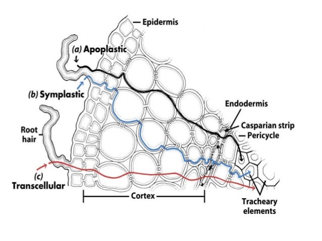
a) Continuous water column:- There are continuous column of  water from the roots through the stem to the leaves. these water column  forms the continuous system through the unthickened areas of the cells called pits.
b) Cohesion or Tensile strength:- Water molecules remain attached with one another  by cohesive forces which is due to hydrogen bonding between adjacent molecules. Therefore, the cohesion force is also called tensile strength. Water molecules will also continue because of adhesive forces between water molecules and walls of tracheary elements.
c) Development of Transpiration Pull:- There is a loss of water from the mesophyll cells of the leaf during transpiration. Because of loss of water these mesophyll cells due to water deficit tends to absorb water from the adjacent cells. Likewise  these adjacent cells after losing the water to  mesophyll cells become water deficit and these cells will absorb water from the tracheids and vessels ( tracheary elements) of the plant. The grater the rate of transpiration the greater will be suction force. With the loss of water from the surface of leaves a pull or tension for is created for water and is known as Transpiration pull.
d) The conducting elements of the plant form a cotinuous system. The transpiration is exerted by living cells of the leaf at the top, throws the water in the xylem in a state of tension. Transpiration pull overcomes the - force of gravity, force of resistance offered by living cells in the roots and resistance offered by xylem channels.


                                                    Path of Water Through the plant

Evidences of transpiration pull :-
i) The rate of water absorption and ascent of sap are almost closely follow the rate of transpiration.
ii) Evaporation of water from a porous pot can produce a tension in a water column.
iii) Dendrography indicates that xylem vessels contract during day time and relax during night. it is due to the transpiration pull.


Wednesday 16 September 2015

Mechanism of Water Absorption in Plants

There are two types of water absorption in plants:-

i) Passive Water Absorption
ii) Active Water Absorption

i) Passive Water Absorption:- This type of water absorption requires very less amount of energy and  takes place because of loss of water from the exposed surface of the plant mainly due to transpiration as a result this creates a tension of low water potential in the xylem cells. Under the influence of this tension water will start getting absorbed from the soil. Water is absorbed due to tension has been proved by various evidences like
a) The absorption of water is almost equal to the rate of transpiration. The pressure of water in the root hairs is -3 to -8 bars and in the soil it is -.1 to -.3 bars. As a result the water of the soil passes into the root hairs.
b) The can be also absorbed by the shoot even in the absence of roots.
c) In tracheary elements water continues to go up due to the tension created by transpiration pull.


ii) Active Water Absorption:- In this type of absorption energy is utilized by the plant and this energy comes from ATP not from the sunlight. Active metabolically living cells are required. There are following possibilities for the active absorption:-
a) Accumulation of the salts and sugars in the trecheary elements of the xylem. Hence DPD of these cells become high results in the absorption of water from the adjacent cells.
b) The cells adjoining the trecheary elements have pumping activities and force the water into trecheary elements. 

Water Absorbing System


Plants have the ability to absorb water from the entire surface like root, stem, leaves, etc. The main source of water for plants is soil. Therefore the root system in plants plays major role in the absorption. Roots are normally very extensive and grow rapidly in the soil. In roots the most efficient part for the absorption of water is root hair zone. This root hair zone has thousands of the minute root hairs which are actively takes part in the absorption. 
There are two pathways in which water passes from root hair zone to xylem inside the roots :-

a) Apoplast Pathway:- In this pathway water moves from root hair zone to xylem through the walls of intervening cells without crossing the  membranes of cells. Therefore in this pathway plants utilizes very less amount of energy because in this pathway there is very less hindrance for passage of water. However this movement of water if interrupted at one place i.e casparian stripes present in endothermal cells.  

Symplast pathway:- This type of pathway is also called transmembrane pathway. In this pathway water has enter into the protoplasm without entering into the vacuoles. The cytoplasm of the adjacent cells are connected through plasmodesmata  ( the bridge between two cells ). For entering into the symplast , water has to pass through the plasmamembrane. This type of absorption involve the expenditure of energy and therfore slower than the apoplastic movements.  


Monday 14 September 2015

Lysosomes

Lysosomes are the small vesicles present in a cell. These small vesicles are containing strong hydrolytic enzymes which are helpful in the intracellular digestion ( the digestion which takes place inside the cell). Lysosomes is a single layered organelle involved in various cellular functions.
                                                                               Lysosomes were first discovered by Belgian scientist, Christian de Duve, in 1955. Lysosomes are present in all animals cells except R.B.C of mammals. In plants and fungi their function is taken over by vacuoles. Enzymes which are present in lysosomes are called acid hydrolases because these enzymes works in the strong acidic medium. The important enzymes are acid phosphatases, sulphatases, proteases, peptidases, nucleases, lipases and carbohydrases.
                                                                                     Lysosomes are mainly originated from plasma membrane by the process of invagination and folding. They are also arise from smooth endoplasmic reticulum or Golgi bodies. The precursors of the enzymes are formed by rough endoplasmic reticulum, then these precursors are transfers to the maturing face of Golgi bodies for maturation. These precursors are then changed into the functional enzymes. These enzymes are then packed in a larger vesicles and these vesicles are when joined with the endosomes are called lysosomes.
                                                                                           For digestion the materials will enter inside the lysosomes where this materials will be acted by the enzymes. Lysomes do not burst in the cytoplasm of the cell to digest any materials.
Polymorphism:- Polymorphism is the term which represents the existence of more than two morphological structures. Depending upon the different structures lysosomes are of four types:-
a) Primary Lysosomes:- These are the newly formed vesicles which are pinched off from Golgi complex. These vesicles when fused with endosomes then will become fully functional. They are small in size.
b) Secondary Lysosomes:- These are also called digestive vacuoles. The food containing phagosomes and lysomes when fuse together will form secondary lysosomes. Digestion occur. The digested food will pass out to the cytoplasm and the lysosomes is left with indigested food.
c) Residual Bodies:- These are the left over or indigested food in the lysosomes. These structures fuse with the plasma membrane to throw out into the external environment called exocytosis or ephagy.
d) Autophagic vacuoles:- These are the fusion of number of primary lysosomes aruond warn out or degenerate intracellular organelles. These organelles are then digested by the strong hydrolytic enzymes of the lysosomes. This is called autophagy or autodigestion.


                                              Functions of Lysosomes
Functions:- 
i) Lysomes are mainly involved in the intracellular as wall as extracellular digestion. The extracellular digestion takes place by the secretion of enzymes by lysosomes outside the cell into external environment.
ii) Lysosomes present in leucocytes helps in the digestion of foreign particles, proteins and microorganisms. In this way they are the main part of immune system.
iii) The lysosomal enzymes present in the acrosome of sperm helps in the breaking limiting membrane of the egg.
iv) They are helpful in the dispose off of useless cells ( aging and dead cells in the body).


Friday 11 September 2015

Golgi Apparatus

Golgi compex is a complex cytoplasmic structure made up of smooth membrane cisternae, a network of tubules with vesicles or vacuoles, which take part in transformation, secretion and production of complex biomolecules. The Golgi complex was discovered by an Italian scientist Camillo Golgi in 1898. The Golgi complex is found in all eukaryotic cells of plants and animals. However, it is absent in prokaryotic cells. In plant cells the Golgi complex consists of number of unconnected structures known as dictyosomes. The shape and size is not always fixed but usually observed that size of Golgi complex is larger in cells which are young and active. The size is small in inactive and old cells.

Structure:- If observed under electron microscope the Golgi complex consists of the following components - i) Cisternae                   ii) Vesicles                 iii) Tubules                 iv) Vacuoles

i) Cisternae:- These are the tubular or flattened fluid filled sacs, separated by intercisternal spaces 200-300 angstom. Each cisterna is a unit membrane structure. Each cisternae consists of two faces. The convex face which is directed towards the plasmalemma is called forming face while the concave face which is directed towards the nucleus is called maturing face. The maturing face is characterised by the presence of large nucleus.

ii) Vesicles:- These are the small drop like structures measuring about 400-800 angstrom in diameter.
   These are associated with convex surface of the Golgi complex. Two types of vesicles smooth and coated vesicles are present.
iii) Tubules:- Theses are structures forming complicated network towards the periphery and maturing face of the cisternae. Tubules have a diameter of 30-50 nm.
iv) Vacuoles:- These are the structures which are present on the maturing surface of the Golgi apparatus. Vacuoles contains an amorphous or granular substances.


Function:- i) The Golgi apparatus is involved in the synthesis of various enzymes which are essential for transformation of membranes.   
ii) Golgi apparatus is actively involved in the secretion through exocytosis or reverse pinocytosis from the cells. It also helps in concentrating and packaging of products of glandular cells.
iii) Proteins released by Rough Endoplasmic Reticulum  and lipids from Smooth Endoplasmis Reticulum reach the cisternae  of the Golgi appartus.
iv) They are also involved in the formation of mucopolysaccarides, pectin, hemicellulose and microfibrills of a cellulose and therfore helps in the synthesis of plant cell wall.


Wednesday 9 September 2015

Endoplasmic Reticulum

It was discovered by Porter (1945) and Thompson (1945). The name was given by Porter in 1953. Endoplasmic reticulum is a 3-dimensional, complicated and interconnected system of membrane-lined channels that run through the cytoplasm. The endoplasmic reticulum is found in all eukaryotic cells except the R.B.Cs of mammals. eggs and embryonic cells. In adipose tissue it is poorly developed. It is extensively developed in the cells which are actively engaged in metabolic activities and protein synthesis e.g. liver cells, cells of pancreas etc.
                                                               
Types:- Depending upon the nature of its membrane, endoplasmic reticulum is of two types, smooth and rough. Endoplasmic reticulum may develop from pre-existing E.R, plasmalemma or nuclear envelop.
Smooth Endoplasmic Reticulum:- It has a smooth membrane which do not bear ribosomes. It is, therfore, also called agranular endoplasmic reticulum. This type of ER is found in cells engaged in the synthesis and storage of glycogen e.g. glycogen storing liver cells, adipose cells, interstitial cells etc. Sphaerosomes are believed to originated from SER.
Rough Endoplasmic Reticulum:- This type of ER bears ribosomes attached to the outer surface of its membrane. RER is, therfore, also called granular endoplasmic reticulum. On account of the presence of ribosomes, the RER engaged in the protein synthesis and secretory activity e.g. plasma cells, pancreatic acinus cells etc.

Structure:- Endoplasmic reticulum consists of membrane lined channels or spaces. The channels contain a fluid called endoplasmic matrix. Endoplasmic reticulum can exist in three forms- Cisternae, Vesicles and Tubules.

Cisternae:- They are flat interconnected sac-like parts of ER which are 40-50 nm in diameter. The cisternae are found in bundles. They occur in the cells actively involved in synthetic activity.
Vesicles:- They are oval and round sacs of 25-500 nm in diameter. They remain isolated in the cytoplasm. The vesicles are also called microsomes. 
Tubules:- They are tube like extensions which may be connected with cisternae or vesicles to form a reticular system. The tubules can be regular or irregular with diameter of 50-100nm.


Functions:-
i) It provides a large surface inside the cell for various physiological activities.
ii) It functions as cytoskeleton by providing mechanical support to cytoplasmic matrix.
iii) Endoplasmic reticulum keeps the various organelles in their position.
iv) Endoplasmis reticulum controls the movement of materials between two adjecent protoplast through plasmodesmata.
v) It provide membranes to nuclear envelop after telophase.
vi) The membranes of ER contains large number of enzymes.
Functions of Rough Endoplasmic Reticulum:- 
i) RER provides a large surface area to ribosomes.
ii) It bears enzymes in the region of pores for modifying polypeptides synthesised by attached ribosomes.e.g. glycosylation.
iii) It provides enzymes precursors for the formation of lysosomes by Golgi Complex.


Thursday 3 September 2015

Plasmolysis

Shrinkage of the protoplast of a cell from its cell wall under the influence of a hypertonic solution is called plasmolysis. Hypertonic solution causes exosmosis Or withdrawal of water from cytoplasm and the the central vacuole of the cell. The size of the cytoplasm as well as central vacuole and hence protoplast becomes reduced. The pressure on the wall is reduced. This is first stage of plasmolysis is called limiting plasmolysis. 
                                          The extra hypertonic solution continues to withdraw water from the central vacuole by exosmosis. Initially the protoplast withdraws itself from the corners. This stage is known as incipient plasmolysis. The hypertonic solution now enters the cell in between the protoplast and the cell wall. Due to continued exosmosis, protoplast shrinks further and withdraws from the cell wall except one or few points. It is known as evident plasmolysis.


Deplasmolysis:- The swelling up of a plasmolysed protoplast under the influence of hypotonic solution or water is called deplasmolysis. It is due to endosmosis. Deplasmolysis is possible only immediately after plasmolysis otherwise the cell protoplast becomes permanently damaged. 

Importance of Plasmolysis;- i) Plasmolysis proves the cell membrane is semipermeable.
ii) It shows that cell wall is elastic as well as permeable.
iii) Osmotic pressure of a cell can be measured by plasmolysis. It will be roughly equivalent to the osmotic pressure of solution.
iv) Plasmolysis can be shown only by living cells.
v) By salting tennis lawns, the weeds can be killed due to permanent plasmolysis.
vi) Salting of pickles, meat and fish kill the spores of fungi and bacteria.


Turgor Pressure

Turgor pressure is the pressure exerted by protoplast upon the cell wall. It occurs due to the osmotic entry of water by endosmosis.
                                               As action and reaction are equal and opposite, therefore, cell wall exerts pressure on protoplast which is called wall pressure. It is denoted by W.P. and is equal to turgor pressure.

Function of Turgor presure:- i) The opening and closing of stomatal pores is regulated by the turgidity of the guard cells.
ii) Turgor pressure provides the mechanical support to the non-woody stems.
iii) It helps in keeping leaves erect and fully expanded. In case of loss of turgidity, the shoots droop down and the leaves show wilting. Wilting is observed in  many plants during summer noon.In wilting the cells of leaves and other softer parts become flaccid due to loss of water. Plants gain their turgidity during night because continued absorption of water from soil, recovery may be partial or it may not occur at all. The latter condition is known as permanent wilting.
iv) Turgor pressure controls the turgor movements, like in the leaf of Mimosa pudica.
v) It helps in cell enlargement.

Diffusion Pressure Deficit

The reduction in the diffusion pressure of water in a solution over its pure state is called diffusion pressure deficit or DPD. It is an older term that was used in place of water potential. Pure water has the maximum diffusion pressure. Diffusion pressure deficit is also called suction pressure. 
                                                                            The term diffusion pressure deficit was given by Meyer (1938). Originally it was described as suction pressure and presently it is replaced by the term water potential. Its value is equal to the osmotic pressure of the solution in a cell minus the wall pressure (= turgor pressure) which opposes the entry of water into it provided the external water is pure.

Interrelationship of OP, TP and DPD:- In a plasmolysed cell the net force with which water enters the cell is equal to its osmotic pressure. As the water enters the cell its turgor pressure increases which acts aganist the osmotic entry of water in a cell.
                                                                                 The net force with which water enters the cell would be equal to the difference of OP and TP.


                                         i.e.            DPD = OP - WP
                                         as               WP = TP
                                     Therfore       DPD = OP - TP

In a flaccid cell
                                             DPD = OP ( as TP = 0 )
In a turgid cell
                                              DPD = 0 ( as OP = TP )


Wednesday 2 September 2015

Plastids

The term plastid was introduced by E. Haeckel in 1886. Plastids are semi-autonomous organelles having DNA and double membrane envelope which store or synthesise various types of organic compounds. Plastids develop from colourless precursors called proplastids. The plastids are large cytoplasmic organelles found in eukaryotic cells. They are absent in all prokaryotes and eukaryotic animal except Euglena and Volvox. On the basis of colour, plastids are of three main types:-

i) Leucoplasts
ii) Chromoplasts
iii) Chloroplasts

i) Leucoplasts:- They are the colourless plastids and do not contain any pigments. They usually perform the function of storage of reserve food material. There are three types of spherical leucoplasts. a) Amyloplasts. They are the starch contaibg leucoplasts. An amyloplast is several times larger than  the original size of leucoplast, e.g. potato tuber, rice, wheat.   b) Elaioplasts. The colourless plastids store fat, e.g. Tube rose.   c) Aleuroplasts, Proteoplasts or Proteinoplasts. The plastids contain protein in the amorphous or crystalloid state, e.g. endosperm cells of Castor.

ii) Chromoplast:- the plastids are yellow or reddish in colour because of the presence of carotenoid pigment. Chlorophylls are absent. Chromoplasts are formed either from leucoplasts or chloroplasts. Change of colour from green to reddish during the ripening of tomato and chilli is due to transformation of chloroplasts to chromoplasts. The orange colour of carrot roots are due to chromoplasts. Some important functions related to chromoplasts are;
a) Chromoplasts provide colours to many flowers for attracting pollinating agents.
b) They provide bright red or orange colour to fruits for attracting animals for dispersal.
c) They are also the site of synthesis of membrane lipids.

iii) Choroplasts:- They are greenish plastids which posses photosynthetic pigments, chlorophylls and carotenoids, and take part in the synthesis of food from the inorganic raw material in the presence of sun light.
             The number of chloroplasts per cell of algae is usually fixed for a species. However different species of different genus may have different number of chloroplasts, e.g. 1 in Spirogyra indica and 16 in S. rectospora. A photosynthetic leaf chlorenchyma cell has 20-40 chloroplasts.
                                                                                           Usually chlororplasts are spherical, oval or discoidal in shape. They are cup-shaped in chlamydomonas and ribbon-shaped in in spirogyra.

Ultrastructure:- A chloroplast has three parts- envelop, matrix and thylakoids.

Chloroplast Envelop:- A chloroplast envelop is made up of two smooth membranes. The outer membrane is more permeable than inner membrane. the outer membrane may be attached to endoplasmic reticulum. At places the inner membrane is connected to thylakoids.
Matrix:-  The ground substance of chloroplast is known as matrix or stroma. It is semifluid colloidal complex that is made up of 50% of soluble proteins.The remaining is DNA, RNA, ribosomes and enzymes. Chloroplast DNA is naked, circular or occasionally linear. DNA makes the chloroplast genetically autonomous because it can both replicate and transcribe to form RNA. Chloroplast ribosomes are 70 S. They resemble the ribosomes of prokaryotes. With the help of ribosomes chloroplast is able to synthesize most of the enzymes required by it. 
Thylakoids :- They are membrane lined flattened sacs which run through out the stroma or matrix of the chloroplast. Since, they take part in photosynthesis. Thylakoids  are thus the structural elements of the chloroplast. In the chloroplast of higher plants, thylakoids are stacked at places to form grana. 40-60 grana may occur in a chloroplast. Each granum has 2-100 thylakoids. Grana are absent in algal chloroplast. The latter are, therefore, agranal. 
                                                                Because of the presence of grana, thylakoids are differentiated into two- granal thylakoids and stroma thylakoids. thylakoids other than grana present in matrix or stroma are called stroma thylakoids. 
                                                                         Thylakoids membranes posses photosynthetic pigments and coupling factor ( involved in ATP synthesis). There are twp photosystems, I and II present on the thylakoid membranes. Photosystem II occurs in appressed parts of granal thylakoids while photosystem I is found in stromal thylakoids and nonappressed parts of granal thylakoids.

Functions:- 
i) Photosynthesis:- Chloroplasts are the centres of photosynthesis and formation of organic compounds from inorganic raw material.
ii) Energy Transduction:- Chloroplasts are able to trap sun energy and change it into chemical energy.
iii) Consumption of carbon Dioxide:- Chloroplasts pick up carbon dioxide and use the same in photosynthesis.
iv) Liberation of Oxygen:- Chloroplasts liberate oxygen which is passed into the atmosphere. This keeps the balance of oxygen constant in the atmosphere. 
v) Storage of starch:- They store starch either temporarily or permanently. 
vi) Photosenstivity:- Chloroplast of some algae provide photosenstivity because of the presence of stigma or eye spot.
vii) Storage of Lipids:- Chloroplast store fat in the form of plastoglobuli.
viii) Formation of chromoplast:- They can be change into the chromoplasts to provide colour to many flowers and fruits.  


Different types of solutions

There are three types of solutions:-

i) Isotonic solution
ii) Hypotonic solution
iii) Hypertonic solution

i) Isotonic solution:- A solution having a concentration such that it neither gain or loses water by osmosis when it is separated by semipermeable membrane from a specific solution eg. 0.9% sodium chloride solution and 5% glucose solution are isotonic.
ii) Hypertonic solution:- The solution having concentration such that it gains water solvent by osmosis across a semipermeable membrane from some other specified solution. When RBCs are placed in a hypertonic solution then exosmosis occurs and RBCs become smaller.
iii) Hypotonic solution:- A solution having a concentration such that it loses water or solvent by osmosis across a semipermeable membrane to some other specified solution. When RBCs placed in hypotonic solution, then endosmosis occurs and RBCs become swollen and spherical.

Tuesday 1 September 2015

Osmosis


Osmosis is defined as the movement of water from its higher chemical potential to its lower chemical potential through semipermeable membrane. The chemical potential of water is also called water potential.                                                          
                                                                          OR

Osmosis is a special type of diffusion that occurs through a semipermeable membrane.


Osmosis is of two types:-     a) Exosmosis                   b) Endosmosis

a) Exosmosis:- It is the outward movement of water from a cell when it is placed in a solution of higher concentration .
b) Endosmosis:- Osmotic inflow of water into a cell when it is placed in a dilute solution having the solute concentration lesser than that of a cell.


Osmotic pressure:- It is the pressure which develops in a osmotically active solution when it is separated from its pure solvent by a semipermeable membrane. Osmotic pressure is also defined as the pressure required to completely stop the entry of water into an osmotically active solution across a semipermeable membrane. It is measured in atomspheres, bars or pascals. The instrument used to measure osmotic pressure is called osmometer.

Significance of osmosis:- i) Osmosis helps in the absorption of water and minerals by roots.
ii) Cell to cell movement of water occur by osmosis.
iii) Opening and closing of stomata are due to osmotic changes in guard cells.
iv) It provides turgidity to guard cells and therefor plant organ.
v) The plant movement like nyctinasty etc. occur due to changes in osmotic pressure.